Your cart is currently empty!
Precisely manufactured for durability and accuracy
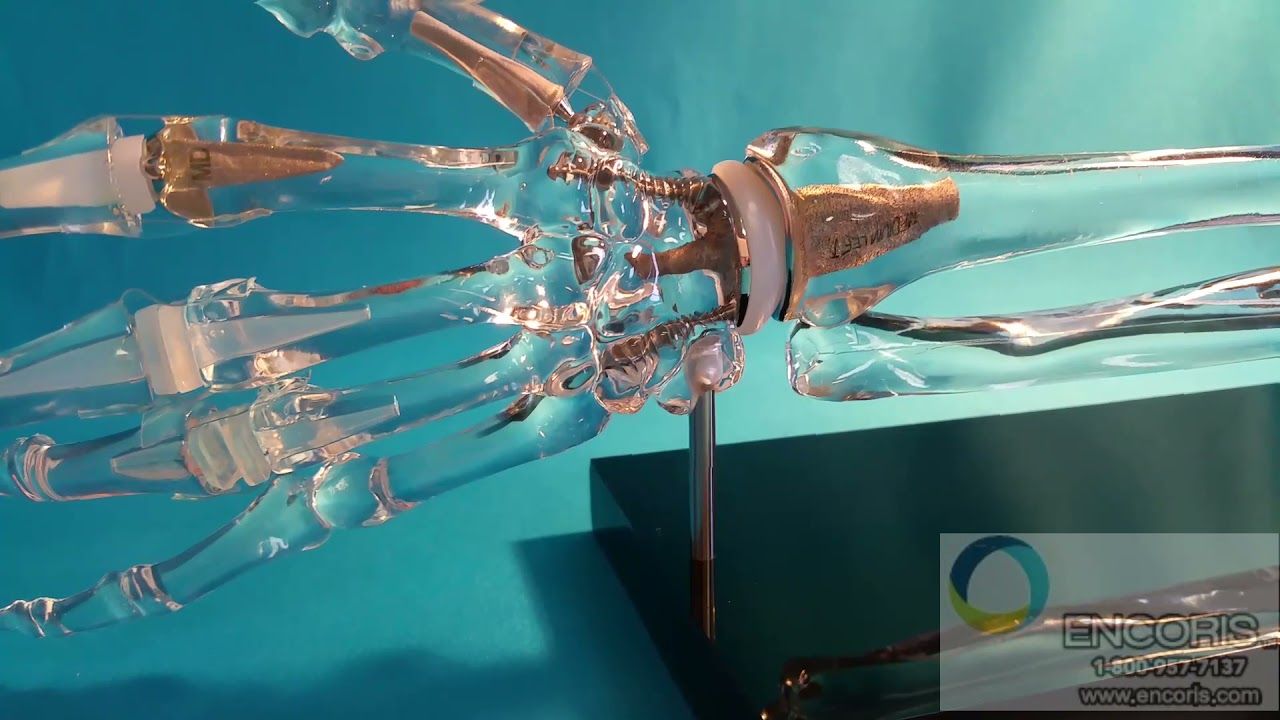
Extremity Models for Medical Training and Demonstration
Our specialized design and manufacturing process streamlines the development of acrylic bone models, no matter how complex the anatomy or implant assembly. We guide clients through each step, often in real time, to ensure every model is accurate from the start and delivered on time with complete confidence.
Our translucent extremity models support clear, in-depth demonstrations of a wide range of orthopedic surgical techniques, including:
Open Reduction/Internal Fixation (ORIF)
Detailed extremity models help illustrate hardware placement and surgical procedures for internal fixation of displaced, comminuted, or unstable fractures of the long bones, metacarpals, metatarsals, and phalanges. We offer highly accurate models of the humerus, radius, ulna, tibia, fibula, femur, foot, and hand—ideal for training and demonstration, especially when introducing procedures to those with limited prior knowledge.
Percutaneous Pinning
Our models are well-suited for demonstrating wire fixation and percutaneous pinning, particularly useful for procedures involving small bones like the phalanges of the hands and feet. The translucent design allows for clear visualization from every angle, making it easy to showcase both the technique and hardware placement.
Tendon Repair
Patients and even healthcare support staff may have limited understanding of tendon repair or tendon transfer procedures, especially in relation to the underlying bony anatomy. Clear anatomical models make it easier to explain tendon positioning and surgical sites with clarity and precision.
Carpal Tunnel Release
Carpal tunnel syndrome is increasingly common in today’s world of repetitive motion and computer use, leading to a growing need for carpal tunnel release procedures. Our hand models are ideal for illustrating the complexities of nerve entrapment and explaining the surgical approach with clarity.
See Surgical Task Trainers or FlexBones

“The team at Encoris is fantastic to work with. They have provided custom models that exceeded expectations. I can’t wait to get them into the hands of our representatives and surgeons!”
Katy Jo J. – MedEd & Marketing Services, Spineology
ACRYLIC EXTREMITY MODELS


